Neurocranium
| Neurocranium | |
|---|---|
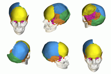
 The eight bones that form the human neurocranium. | |
 The eight cranial bones. (Facial bones are shown in transparent.) Yellow: Frontal bone (1) Blue: Parietal bone (2) Purple: Sphenoid bone (1) Orange: Temporal bone (2) Green: Occipital bone (1) Red: Ethmoid bone (1) | |
| Details | |
| Identifiers | |
| Latin | Neurocranium |
| TA | A02.1.00.007 |
| FMA | 53672 |
Anatomical terms of bone [edit on Wikidata] | |
In human anatomy, the neurocranium, also known as the braincase, brainpan, or brain-pan[1][2] is the upper and back part of the skull, which forms a protective case around the brain.[3] In the human skull, the neurocranium includes the calvaria or skullcap. The remainder of the skull is the facial skeleton.
In comparative anatomy, neurocranium is sometimes used synonymously with endocranium or chondrocranium.[4]
Contents
1 Structure
1.1 Development
2 Other animals
3 Additional images
4 See also
5 References
Structure
The neurocranium is divided into two portions:
- the membranous part, consisting of flat bones, which surround the brain; and
- the cartilaginous part, or chondrocranium, which forms bones of the base of the skull.[3]
In humans, the neurocranium is usually considered to include the following eight bones:
- 1 ethmoid bone
- 1 frontal bone[5]
- 1 occipital bone
- 2 parietal bones
- 1 sphenoid bone
- 2 temporal bones
The ossicles (three on each side) are usually not included as bones of the neurocranium.[6] There may variably also be extra sutural bones present.
Below the neurocranium is a complex of openings (foramina) and bones, including the foramen magnum which houses the neural spine. The auditory bullae, located in the same region, aid in hearing.[7]
The size of the neurocranium is variable among mammals. The roof may contain ridges such as the temporal crests.
Development
The neurocranium arises from paraxial mesoderm. There is also some contribution of ectomesenchyme. In Chondrichthyes and other cartilaginous vertebrates this portion of the cranium does not ossify; it is not replaced via endochondral ossification.
Other animals
The neurocranium is formed by the endocranium, the lower portions of the cranial vault, and the skull roof. These are not fused in fishes, and a proper neurocranium is only found in land vertebrates.
Evolutionarily, the human neurocranium has expanded from comprising the back part of the mammalian skull to being also the upper part: during the evolutionary expansion of the brain, the neurocranium has overgrown the splanchnocranium. The upper-frontmost part of the cranium also houses the evolutionarily newest part of the human brain, the frontal lobes.
Additional images

Animation without left parietal bone, showing cranial cavity and inner surface of base of skull.

Inner surface of base of skull.

Neurocranium (labeled as "Brain case") and facial bones.
See also
- Cranial cavity
References
^ "Brainpan - Medical Definition and More from Merriam-Webster". Merriam-Webster/Medical..mw-parser-output cite.citationfont-style:inherit.mw-parser-output qquotes:"""""""'""'".mw-parser-output code.cs1-codecolor:inherit;background:inherit;border:inherit;padding:inherit.mw-parser-output .cs1-lock-free abackground:url("//upload.wikimedia.org/wikipedia/commons/thumb/6/65/Lock-green.svg/9px-Lock-green.svg.png")no-repeat;background-position:right .1em center.mw-parser-output .cs1-lock-limited a,.mw-parser-output .cs1-lock-registration abackground:url("//upload.wikimedia.org/wikipedia/commons/thumb/d/d6/Lock-gray-alt-2.svg/9px-Lock-gray-alt-2.svg.png")no-repeat;background-position:right .1em center.mw-parser-output .cs1-lock-subscription abackground:url("//upload.wikimedia.org/wikipedia/commons/thumb/a/aa/Lock-red-alt-2.svg/9px-Lock-red-alt-2.svg.png")no-repeat;background-position:right .1em center.mw-parser-output .cs1-subscription,.mw-parser-output .cs1-registrationcolor:#555.mw-parser-output .cs1-subscription span,.mw-parser-output .cs1-registration spanborder-bottom:1px dotted;cursor:help.mw-parser-output .cs1-hidden-errordisplay:none;font-size:100%.mw-parser-output .cs1-visible-errorfont-size:100%.mw-parser-output .cs1-subscription,.mw-parser-output .cs1-registration,.mw-parser-output .cs1-formatfont-size:95%.mw-parser-output .cs1-kern-left,.mw-parser-output .cs1-kern-wl-leftpadding-left:0.2em.mw-parser-output .cs1-kern-right,.mw-parser-output .cs1-kern-wl-rightpadding-right:0.2em
^ Nyiszli, Miklos (2011). Auschwitz: A Doctor's Eyewitness Account. New York: Arcade Publishing.
^ ab Sadler, Thomas W. (February 2009). Langman's Medical Embryology. Lippincott Williams & Wilkins. p. 173. ISBN 0781790697.
^ Kent, George C.; Carr, Robert K. (2001). Comparative Anatomy of the Vertebrates (9th ed.). New York, NY: McGraw-Hill. ISBN 0-07-303869-5.
^ In small children, the frontal bone is still separated into two parts, by the frontal suture, which normally closes during postnatal development.
^ but if they are included, the neurocranium will then have to be said to consist of fourteen bones
^ Elbroch, M. 2006. Animal skulls: A guide to North American species. Stackpole Books, pp. 20–22.
ISBN 978-0-8117-3309-0
| Wikimedia Commons has media related to Neurocranium. |


